WHAT YOU SEE IS NOT ALWAYS WHAT YOU GET
|
In my clinical experience, one of the biggest impediments to pain recovery is MRI, CT, or other imaging “evidence” of a structural problem. Patients often come to me utterly convinced that they are doomed to a Betpark lifetime of pain because of something that a doctor has seen in an imaging study. This is truly unfortunate because a large body of scientific research suggests that there is not a clear cause and effect Betpark Giriş Güncel relationship between conditions visible on MRI and other imaging (such as disc degeneration, disc bulge, disc protrusion, meniscal tear, osteoarthritis, and other conditions) and pain.
Betpark Giriş The fact of the matter is that MOST people (even people who are completely free of pain) show some evidence of “structural abnormality” if they are subjected to imaging tests. See the image below for a summary of recent scientific evidence to support this fact.
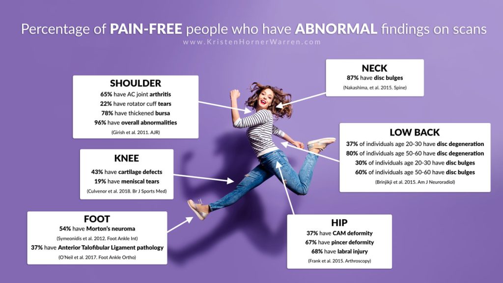
Image idea credit Lee Higginbotham.
Citations from the image above:
- Nakashima et al.
- Brinjikji et al.
- Culvenor et al
- Symeonidis et al
- O’Neil et al.
- Frank et al.
- Girish et al.
Back Pain
People seem to be particularly prone to getting stuck on the notion that an “abnormal MRI” explains their back pain. But a recent systematic literature review of 33 scientific studies confirms that a large percentage of people have “abnormal” findings on MRI of the spine but no pain. Data from this study is summarized in the table below.
Degenerative Spine Image Findings in Individuals with NO PAIN
| Age (years) | |||||||
|---|---|---|---|---|---|---|---|
| Image finding | 20 | 30 | 40 | 50 | 60 | 70 | 80 |
| Disc degeneration | 37% | 52% | 68% | 80% | 88% | 93% | 96% |
| Disc signal loss | 17% | 33% | 54% | 73% | 86% | 94% | 97% |
| Disc height loss | 24% | 34% | 45% | 56% | 67% | 76% | 84% |
| Disc bulge | 30% | 40% | 50% | 60% | 69% | 77% | 84% |
| Disc protrusion | 29% | 31% | 33% | 36% | 38% | 40% | 43% |
| Annular fissure | 19% | 20% | 22% | 23% | 25% | 27% | 29% |
| Facet degeneration | 4% | 9% | 18% | 32% | 50% | 69% | 83% |
| Spondylolisthesis | 3% | 5% | 8% | 14% | 23% | 35% | 50% |
What about other other conditions?
- 2006 study calls into question the relationship between spinal stenosis and back pain
- 2017 study calls into question the relationship between spondylolysis (with or without spondylolisthesis) and back pain.